
Figure 6.
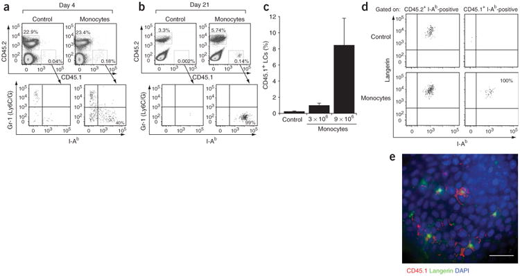
Adoptive transfer of Gr-1hi monocytes in vivo. (a,b) Top, percent propidium iodide–negative–gated epidermal cells that are CD45.1+ or CD45.2+ in mice injected with monocytes or control (I-Ab+ CD11c+) populations. Bottom, Gr-1 and I-Ab expression patterns of gated epidermal CD45.1+ cells in mice injected with monocytes or control (I-Ab+ CD11c+) populations, 5 d (a) or 21 d (b) after exposure to ultraviolet irradiation. Results are representative of three separate experiments. (c) Percent CD45.1+ LCs derived from adoptively transferred CD45.1+ cells among total LCs. Data represent percent LCs derived from 3 × 10⁶ or 9 × 10⁶ monocytes or 9 × 10⁶ enriched bone marrow I-Ab+CD11c+ cells (Control) and are from one representative experiment (n = 3). (d) Langerin expression pattern of gated CD45.2+ I-Ab+ and CD45.1+ I-Ab+ epidermal cells in mice 6 weeks after adoptive transfer of CD45.1+ monocytes. Results are representative of two individual mice. (e) Epidermal sheet stained with CD45.1 (red), langerin (green) and DAPI (blue). Epidermis is from a CD45.2+ mouse, 6 weeks after adoptive transfer of 9 × 10⁶ CD45.1+ monocytes. Scale bar, 10 μm. Results are representative of two individual mice.