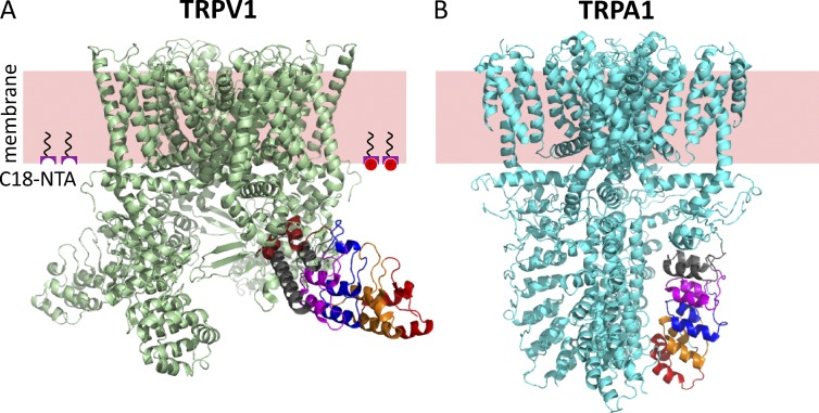
Figure 1.
Structural divergence in ankyrin repeat domains of TRPV1 and TRPA1. (A) Structure of TRPV1 (3J5P) with ankyrin repeats 1–6 labeled in red, orange, blue, magenta, gray, and brown. The pink slab represents the approximate location of the plasma membrane, and the moieties at the intracellular surface represent C18-NTA partitioned into the membrane. Co2+ is represented by red circles. (B) Structure of TRPA1 (3J9P) with ankyrin repeats 12–16 shown in red, orange, blue, magenta, and gray.

