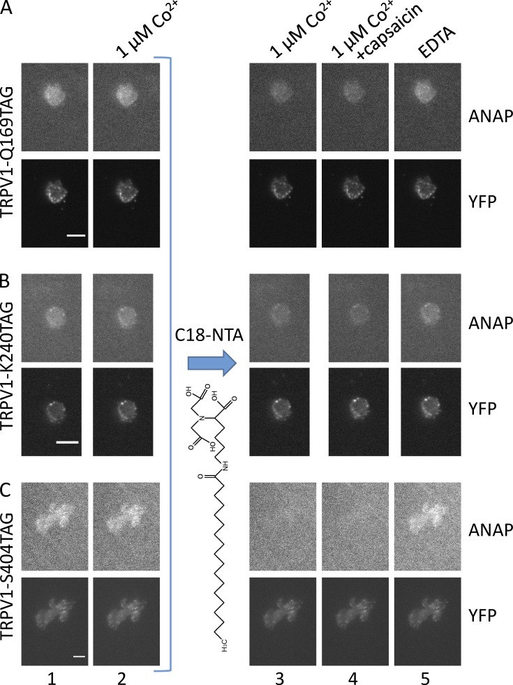
Figure 9.
tmFRET occurs between L-ANAP incorporated into TRPV1 and Co2+ bound to C18-NTA in the membrane. (A–C) ANAP and YFP fluorescence from unroofed cells cotransfected with pANAP and TRPV1-Q169TAG-YFP (A), TRPV1-K240TAG-YFP (B), and TRPV1-S404TAG-YFP (C) and cultured with L-ANAP-ME in the medium. Images were acquired with an ANAP-appropriate cube and a YFP-appropriate cube as indicated. For each field, the same camera settings and lookup table were used for all images in the same color channel. Epifluorescence images were acquired in the sequence shown starting with the initial fluorescence with no added Co2+ (column 1) and then in the presence of 1 µM Co2+ (column 2). Subsequently, the unroofed cells were incubated in C18-NTA as described in Materials and methods, and images were acquired in the presence of 1 µM Co2+ (column 3), 1 µM Co2+ with capsaicin (column 4), and EDTA (column 5). Scale bars for all panels are 10 μm.

