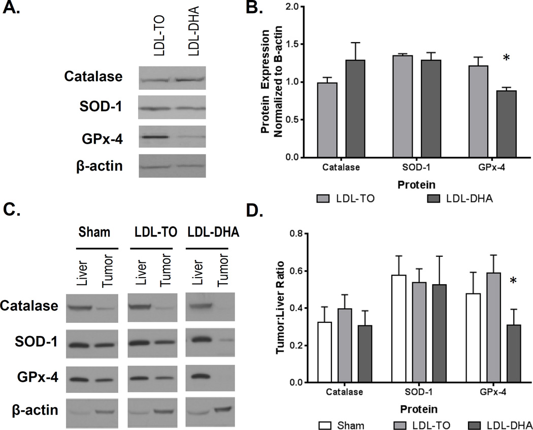
Figure 6. In vitro and in vivo protein expression of catalase, SOD-1 and GPx-4 following LDL nanoparticle treatments.
(A, B) Protein expression levels of enzyme antioxidant 72 hours after LDL nanoparticle (40 µM) treatments in H4IIE cells. Representative blots and quantifications are shown. The data are expressed relative to β-actin expression (mean ± SEM) for each treatment group. *, P ≤ 0.05 (C,D) Protein expression levels of enzyme antioxidants in rat liver and H4IIE tumor 72 hours after Sham and HAI treatment of LDL nanoparticles (2 mg/kg). Representative blots and quantifications are shown. The data are expressed relative to β-actin expression as a ratio of tumor to liver (mean ± SEM) for each treatment group. *, P ≤ 0.05

