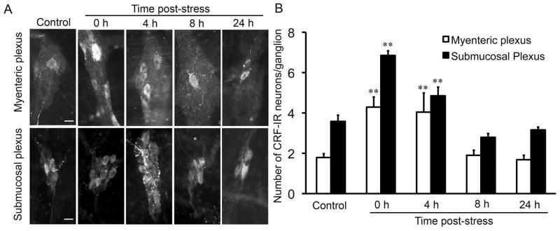
Fig. 3.
Time course of the effects of partial restraint stress on the number of CRF-immunoreactive neurons in the enteric nervous system of the rat colon. Immunofluorescence staining was used to determine the number of CRF-immunoreactive (CRF-IR) neurons in the myenteric and submucosal plexuses of the rat colon. (A) Representative images showing the effects of PRS on the number of CRF-IR neurons in the enteric nervous system. In control rats, only one or two CRF-IR neurons were found in each myenteric or submucosal ganglion. Immediately after PRS, the number of CRF-IR neurons/ganglion increased significantly. The increase in CRF-IR neurons lasted for 4 h and returned to normal levels after 8 h. (B) Bar graph showing the effects of PRS on CRF-IR neurons. Scale bar = 20 μm. N = 5–8/group; ** P < 0.01 compared to control.

