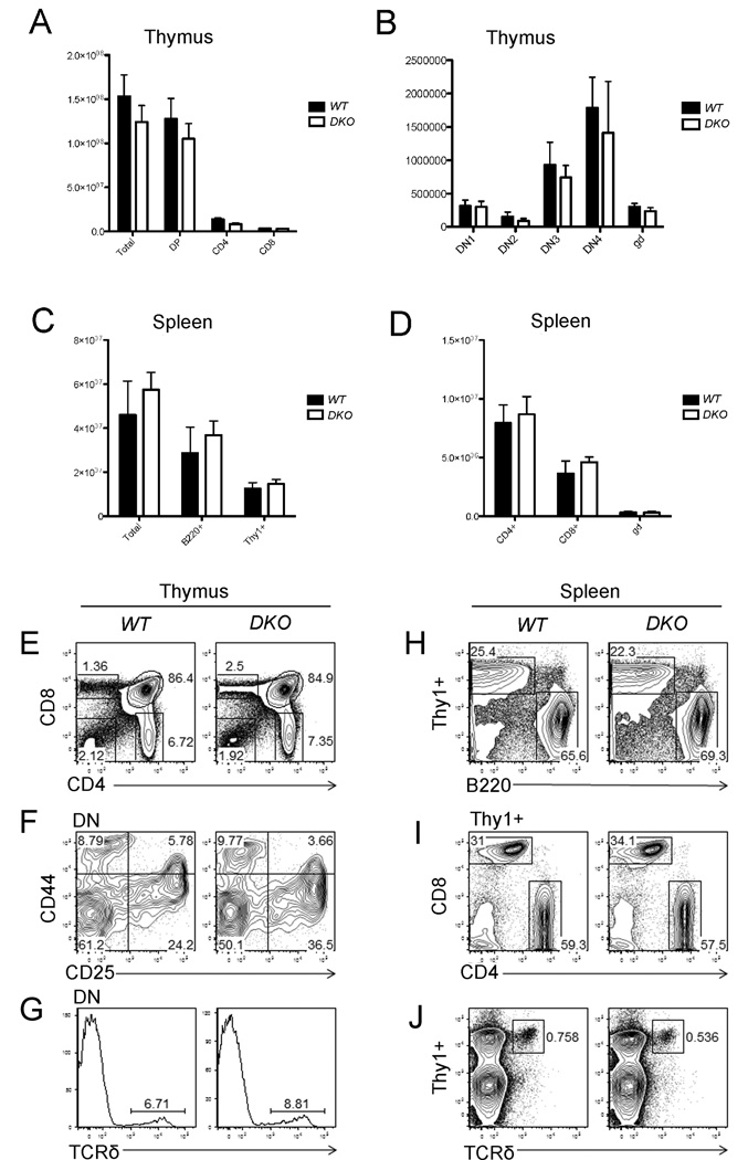
Figure 4.
Co-deletion of Appl1 and Appl2 do not affect T-cell development. WT and Appl DKO mice at 6 weeks of age were sacrificed to collect thymus (A, B) and spleen cells (C, D). Cells were stained with fluorescence-labeled anti-CD4, CD8, CD44, CD25, Thy1 and B220 antibodies, and then counted using multicolor flow cytometry. (A) CD4+, CD8+ and CD4+CD8+ (DP) cells in thymus. (B) Distribution of various stages of CD4/CD8 double-negative (DN) cells in thymus of WT and Appl DKO mice. gd = γδ T cells. (C) Thy1+ and B220+ population in spleen. (D) Mature CD4+ and CD8+ T-cells in spleen. (E) Percentages of CD4+, CD8+ and CD4+CD8+ and CD4-CD8- populations in thymus of WT and DKO mice. (F) Distribution of CD44+, CD25+, CD44+CD25+, and CD44-CD25- cells. (G) TCR δ population in thymus cells from WT and DKO mice. (H, I, J) Percentages of Thy1+ and B220+ populations, mature CD4+ or CD8+ T-cells, and TCR δ T cells in spleen, respectively.

