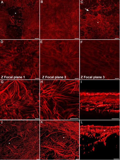
Figure 4. Multiple glial blooms coalesced to form continuous glial membranes.
GFAP labeling of wholemount retinas demonstrates large glial membranes. (A) This tiled image of a posterior pole shows two large membranes merging above a normal astrocyte pattern (asterisk). (B) A continuous glial membrane covers the entire area of this tiled imaged. (C) Connections between blooms (arrow) create this larger glial membrane (asterisk). (D) Higher magnification demonstrates the density of these structures. (E) In the focal plane below “D”, numerous GFAP+ processes are present. (F) Astrocytes in the retina below are tortuous with misaligned processes. (G) In another membrane, thick, GFAP+ processes arborize into smaller processes which connect with one another. (H) Many smaller structures within this membrane appear to be individual cells. Long processes extend between these glial cells and into the retina. (I) A digital cross sectional view demonstrates the multiple layers of GFAP+ cells. The connections between the top layer (which lies in the vitreous) and astrocytes below can be observed. (J) The complexity of membranes is also shown in this 3D rendering. The astrocytes in an area lacking membrane (asterisk) are visible. (K) A side view of this membrane shows the astrocyte layer below (asterisks). (L) A cross sectional view demonstrates the astrocytes (asterisk) below the glial membrane. Scale bars indicate: A-C: 500 μm; D-H: 50 μm; I: 10 μm, and J-L: 20 μm.

