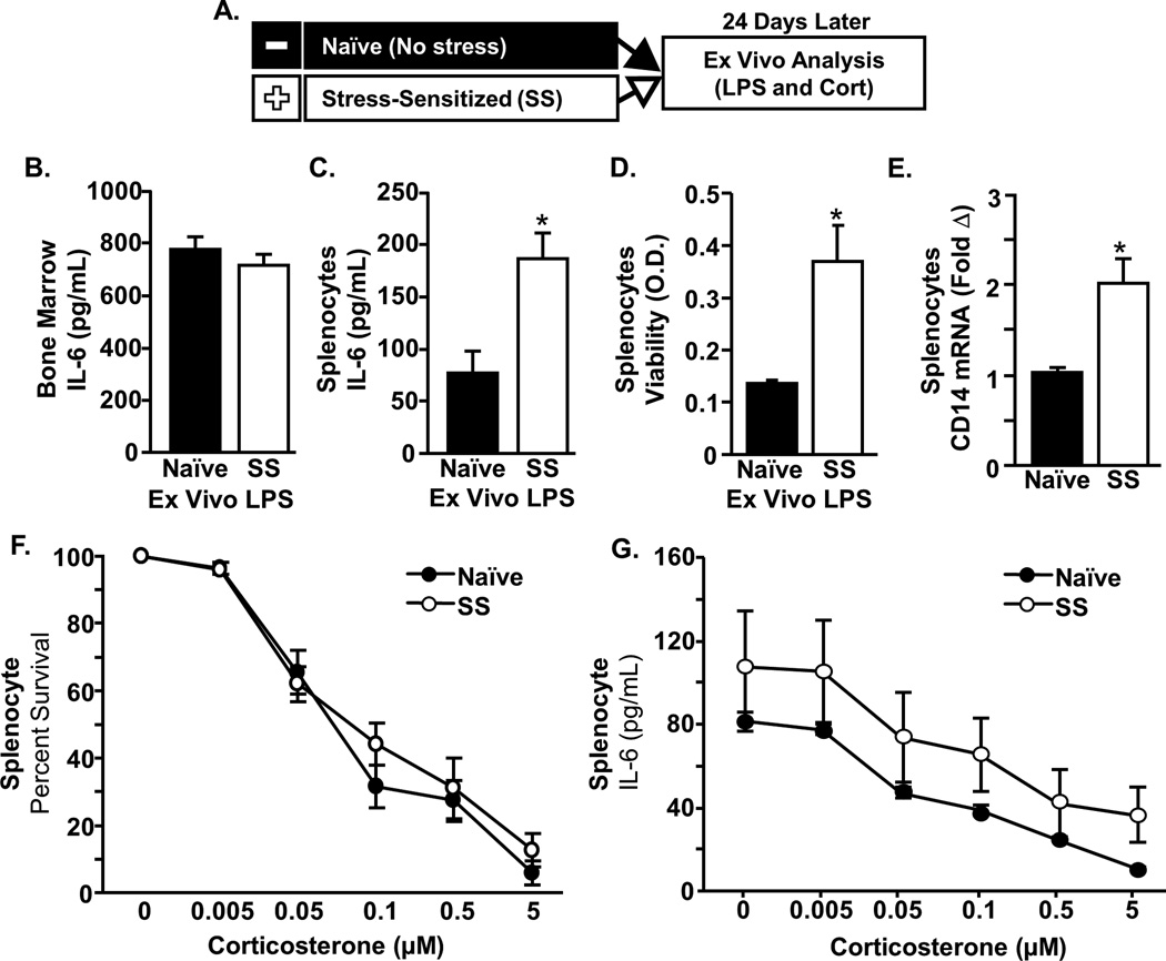
Figure 4. Stress-sensitization resulted in the accumulation of primed splenocytes with an enhanced response to ex vivo mitogen challenge.
A) Male C57BL/6 mice were stress-sensitized (SS) by 6 repeated cycles of social defeat or left undisturbed as controls (Naïve). Twenty four days later, spleen and bone marrow (BM) cells were cultured ex vivo in the presence of lipopolysaccharide (LPS) and corticosterone (cort). IL-6 protein was determined in the cell supernatants collected 18 h after LPS in BM and Splenocytes. B) IL-6 secretion was similar between groups in bone marrow cells. C) The LPS induced IL-6 secretion was higher in splenocytes cultured from SS mice compared to naïve mice (p<0.01). D) Cell viability of LPS-stimulated splenocytes was increased in SS mice (p<0.05). E) mRNA expression of CD14 in SS splenocytes was higher than naïve (p<0.01). Next, ex vivo cultures from spleen were stimulated with LPS for 48 h in the presence of increasing concentrations of corticosterone and cell viability and IL-6 concentrations were determined. F) There was a main effect of corticosterone on viability (F5,36=6.84, p<0.0001) and G) on IL-6 production F5,36=4.42, p<0.005) that was independent of stress. F&G) There was also a main effect of stress on viability (F1,36=9.63, p<0.005) and IL-6 production (F1,36=4.69, p<0.05). Bars represent the mean ± SEM. Means with asterisk (*) are significantly different from CON (p<0.05) according to F-protected post hoc analysis.

