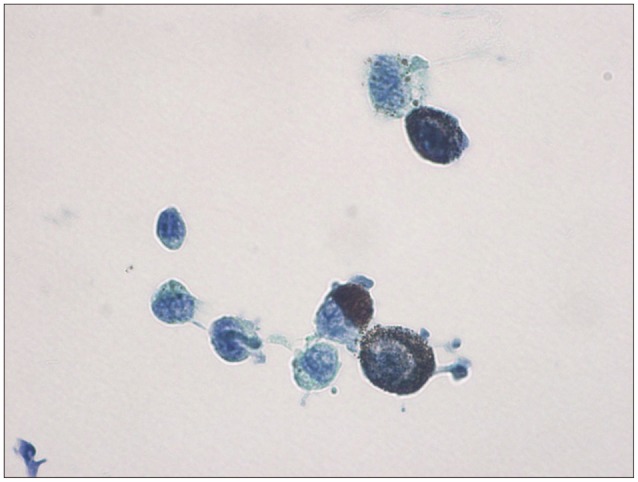
Fig. 3. Pathological findings shows pleomorphic cells with abundant cytoplasm, abundant intracytoplasmic melanin pigments, nuclear pleomorphism, and prominent nucleoli (Papanicolaou stain, ×1000). These tumor cells showed strong immunoreactivity to HMB-45, S-100, and Melan-A on the immunohistochemical study.

