
Figure 1.
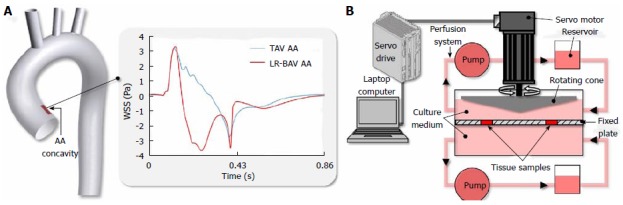
Ex vivo methodology. A: Temporal wall shear stress (WSS) signals captured computationally in the concavity of the tricuspid aortic valve (TAV) ascending aorta (AA) and left-right bicuspid aortic valve (LR-BAV) AA (adapted from Cao et al[28]); B: Shear stress bioreactor used to condition porcine AA tissue to TAV AA and BAV AA WSS.