. 2015 Oct 29;4:e10721. doi: 10.7554/eLife.10721

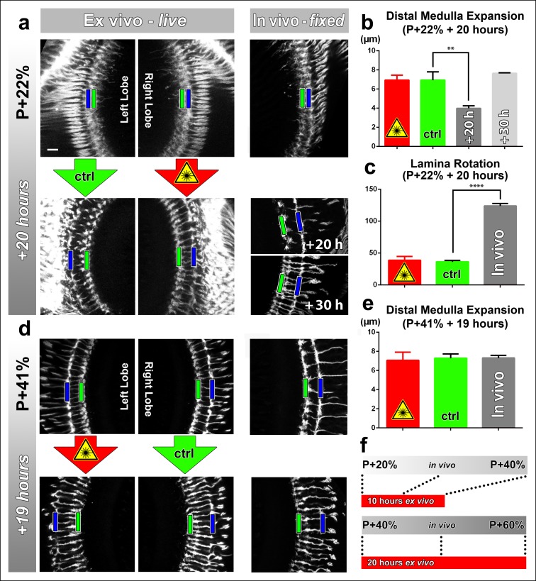
Figure 2. Effects of culture conditions and laser scanning on the optic lobe development ex vivo.

Two-photon imaging of the medulla was performed with brains cultured at P + 22% for 20 hr (a) and P + 41% for 19 hr (d) all photoreceptors express CD4-tdGFP. For each experiment one image stack was acquired containing both optic lobes of a brain. Next, only one of the lobes was scanned every 30 min. Finally, another stack was acquired with both lobes. Different brains aged in parallel in pupae have been dissected as in vivo controls. (b) Quantification of the layer distance increase in P + 22% cultures. The distance between R8 (green rectangles in a,d) and R7 (blue rectangles) layers increase identically in scanned and unscanned ex vivo lobes, but higher than the in vivo control (p = 0.0036, n = 3). (c) Quantification of the change in the angle between the planes of posterior lamina and the anterior medulla. Ex vivo lobes rotate similarly but slower than in vivo controls (p <0.0001, n = 3). (e) Quantification of the layer distance increase in P + 41% cultures. All groups show a similar increase in the distance between R8 temporary layer and R7 terminals. Error bars depict SEM. (f) Calibration of the developmental speed in culture to in vivo development, based on distal medulla expansion. Scale bars, 10 μm.

DOI: http://dx.doi.org/10.7554/eLife.10721.007

Figure 2.

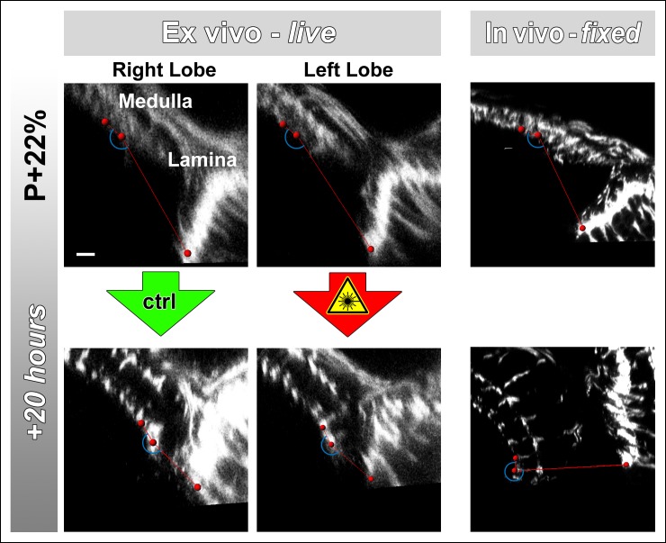
Figure 2—figure supplement 1. Lamina rotation is incomplete ex vivo.

Figure 2—figure supplement 1.

Two-photon imaging of the medulla was performed with brains cultured at P + 22% for 20 hr, all photoreceptors express CD4-tdGFP. Continuously scanned ex vivo culture, unscanned control optic lobe and in vivo (fixed) control experiments were done as described in Figure 2. The angles (blue arches) between the planes of posterior lamina and anterior medulla have been measured for the start and end points of each culture as well as the corresponding in vivo controls; and plotted in Figure 2c. Scale bar, 10 μm.
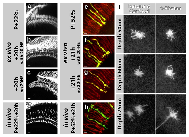
Figure 2—figure supplement 2. Effects of 20-Hydroxyecdysone (20-HE) and type of microscope on imaging in the culture chamber.

Figure 2—figure supplement 2.

(a-h) 20-HE is required for early but detrimental to late pupal development in the optic lobe. (a-d) All photoreceptors were labeled with CD4-tdGFP. Cultures were set-up at P + 22% (a), with (b) or without (c) 20-HE in the culture media. Parallel developed pupae were dissected and imaged at the end of cultures as in vivo controls (d). R7-R8 layer separation in the medulla was impaired in cultures without 20-HE compared to in vivo controls or cultures with 20-HE. Scale bars, 10 μm. (e-h) All photoreceptors were labeled with td-Tomato and R7 cells were sparsely labeled with CD4-tdGFP using GMR-FLP through MARCM. Cultures were set-up at P + 22% (e), with (f) or without (g) 20-HE in the culture media. Parallel developed pupae were dissected and imaged at the end of cultures as in vivo controls (h). R7 axons that developed in the presence of 20-HE showed excessive filopodial formations on their terminals compared in vivo controls or the cultures without 20-HE. Scale bars, 4 μm. (i) Comparison of resonant confocal and 2-photon microscopy signal stengths in the imaging culture chambes. R7 cells were sparsely labeled with CD4-tdGFP using GMR-FLP through MARCM. Individual R7 growth cones were imaged in the culture chamber at P + 30%. Images were acquired with a Leica TCS SP5 confocal microscope with a resonant scanner or a Zeiss LSM 780 multiphoton microscope at various depths from the coverslip. The confocal signal reduces below 60 μm compared to the 2-photon signal.