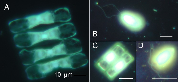
Fig 10. Images by epifluorescence microscopy of brine samples.
Organisms are stained with acridine orange and DAPI according to a dual-staining technique [41] and showed an assemblage of diatoms (A, C) and flagellates (B, D) from Malene Bay (A, B) and Kobbefjord (C, D). All scale bars are 10 μm. From Malene Bay: (A) A diatom, probably Navicula vanhoeffenii and (B) a flagellate, possibly Chlamydomonas or Telonemia; from Kobbefjord: (C) a diatom, probably Fragilariopsis oceanica, and (D) a flagellate, Pyraminmonas sp. (http://westerndiatoms.colorado.edu/) [46] [47].

