Fig. 1.
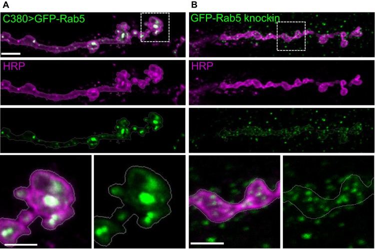
Overexpression of the endosomal marker GFP-Rab5 changes its localization and distribution pattern. (A) C380-Gal4-driven UAS-GFP-Rab5 localizes to large punctate compartments at neuronal termini (outlined by HRP staining) that innervate larval muscles, and appear quite different from (B) endogenously expressed GFP-Rab5 (Fabrowski et al., 2013) compartments, which are smaller in size and fairly uniformly distributed. In GFP knock-in animals, GFP-Rab5 is also visible in the postsynaptic muscle tissue, reflecting its endogenous expression pattern. Muscle 6/7 NMJ is shown from segment A3. Scale bars are 5 µm for top panels and 2.5 µm for magnified bottom panels.

