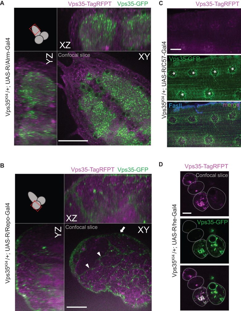
Fig. 4.
Vps35 tagging in astrocytes, pan-glial populations, larval muscles and larval hemocytes. (A) Astrocyte-specific Vps35 GFP tagging. YZ and XZ single confocal planes demonstrating the subcellular distribution of Vps35-GFP throughout the larval astrocyte population, including their processes infiltrating the neuropil. Scale bar is 40 µm. (B) Repo-Gal4-mediated pan-glial tag conversion reveals surface glial (arrow) and cortex glial (arrowheads) expression of Vps35 in a single confocal plane through the lobe of the larval CNS. Scale bar is 40 µm. (C) Larval muscle-specific Vps35 GFP tagging. Maximum intensity projection of a confocal Z-stack through a larval muscle cell M6 (which are multi-nucleate, marked by stars) that expresses Rippase with the muscle driver C57-Gal4. Vps35-GFP is present most prominently around the muscle nuclei though it can be seen in small punctate structures throughout the muscle cell. Synaptic termini were stained with FasII. Scale bar is 25 µm. (D) Hemocyte-specific Vps35 GFP tagging. Single confocal slice through a cluster of four third instar larval hemocytes from Vps35KI4 knock-in animals that express Rippase with the hemese-Gal4 driver in the majority of hemocytes (Zettervall et al., 2004). The top two cells express Vps35-TagRFPT almost exclusively, with only very low level of Vps35-GFP signal detectable, indicating that Vps35-GFP translation has not progressed long enough for the signal to become readily visible. The other hemocytes accumulated varying amounts of Vps35-GFP depending on their cellular birth date and the relative timing of R-mediated conversion. Scale bar is 5 µm.

