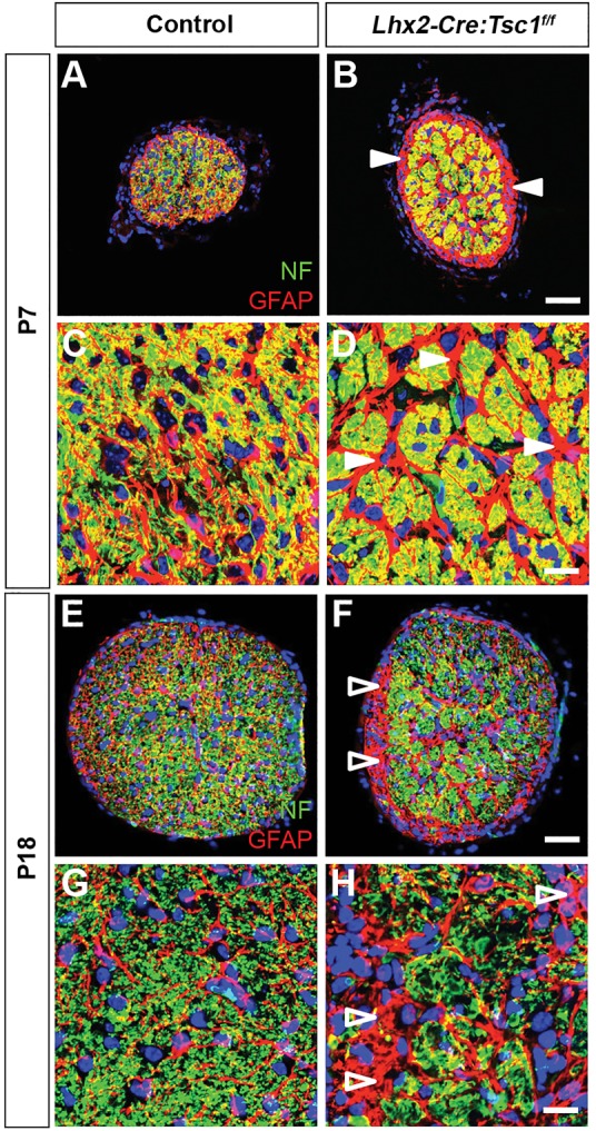
Fig. 6.

Conditional deletion of Tsc1 leads to reactive gliosis and changes in optic nerve morphology. (A-D) Transverse sections of the ON at P7 demonstrate a defined network of GFAP+ astrocytes that enmesh the NF+ RGC axons in control mice (A,C). In contrast, regionalised areas of reactive gliosis are clearly visible in the ON of Lhx2-Cre:Tsc1f/f mice (B,D, arrowheads). (E-H) Transverse sections of the ON at P18 demonstrate a defined network of GFAP+ astrocytes that enmesh the NF+ RGC axons in control mice (E,G). In contrast, axon loss is clearly visible in regionalised areas of the ON from Lhx2-Cre:Tsc1f/f mice. Also evident are cross-sectional areas that are significantly enriched in GFAP+ astrocytes and completely devoid of NF+ RGC axons (F,H, arrowheads). Scale bars: (A,B;E,F) 50 µm; (C,D;G,H) 25 µm. Abbreviations: ON, optic nerve; GFAP, glial fibrillary acidic protein; NF, neurofilament; RGC, retinal ganglion cell.
