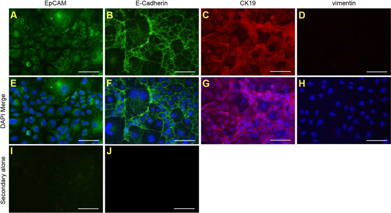
Fig. 3.
Cell lines derived from mice fed a CDE diet express LPC markers. The LPC line, BMOL3, was derived from a mouse fed a 67% CDE diet. Immunofluorescence was used to detect known LPC markers [EpCAM (A,E), E-cadherin (B,F) and CK19 (C,G)] and mesenchymal marker vimentin (D,H). Representative images are shown without (A-D) and with (E-H) DAPI staining. Control staining with either Alexa-Fluor-488 goat-anti-rabbit or -594 goat-anti-rat alone are shown (I,J). Scale bars: 100 µm.

