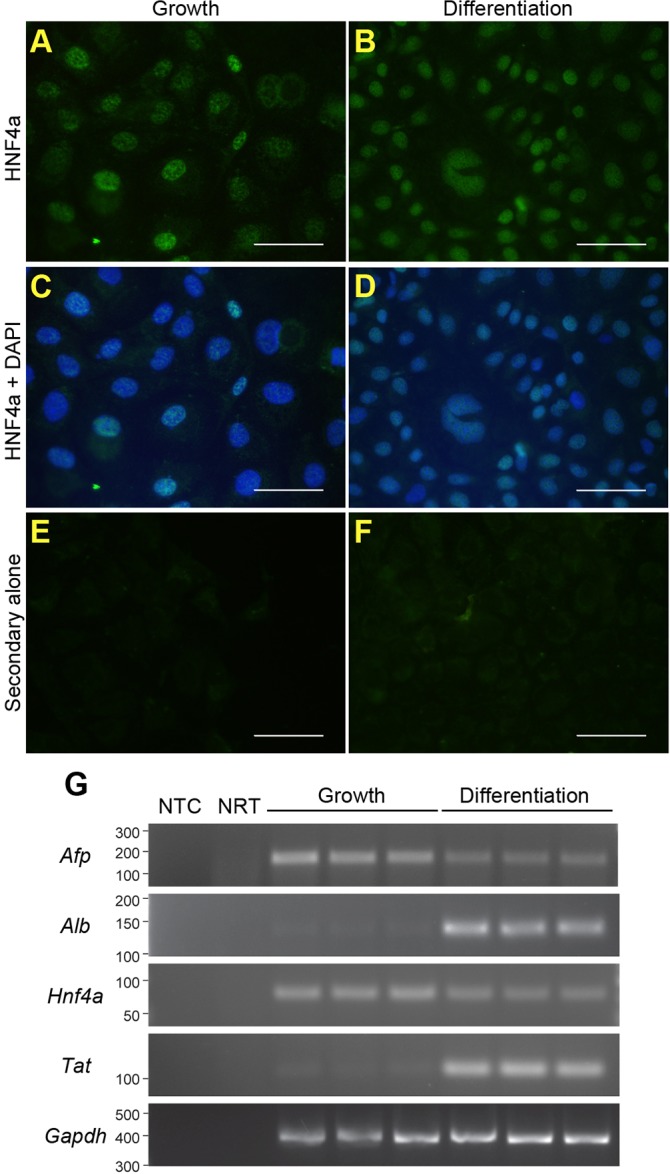
Fig. 4.

BMOL3 LPCs can differentiate towards the hepatocyte lineage. BMOL3 cells were treated to differentiate them towards hepatocytes. Proliferating (A,C,E) and differentiated (B,D,F) LPCs were stained for HNF4α (A,B). Images are shown without (A,B) and with (C,D) DAPI staining. Control staining with Alexa-Fluor-488 rabbit-anti-goat alone is shown (E,F). Scale bars: 50 µm. RT-PCR was used to determine abundance of the hepatocyte markers Afp, Alb, Hnf4α and Tat. Gapdh was used as a loading control. Non-template (NTC) and no reverse transcriptase enzyme (NRT) were included as controls.
