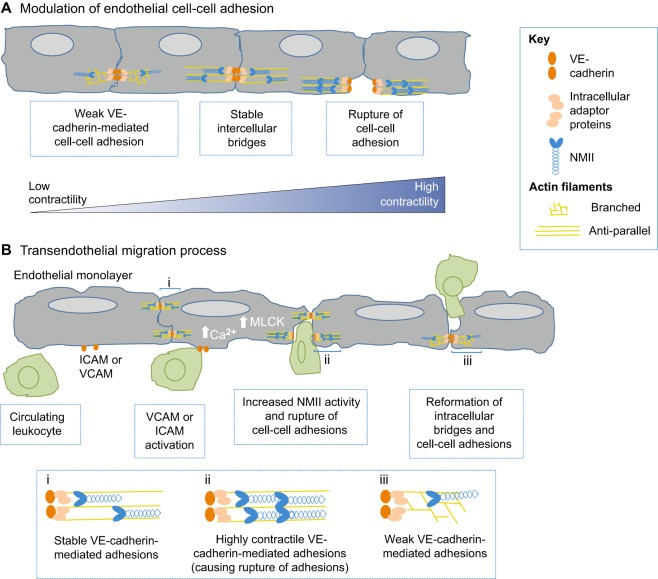
Fig. 4.
NMII regulates vascular biology and disease. (A) NMII remodels endothelial adherens junctions. VE-cadherin (orange ovals) mediates intercellular links between neighboring cells. Intracellularly, catenins (beige ovals) link VE-cadherin with the actin cytoskeleton (yellow lines). Myosin (blue) generates forces that stabilize intercellular bridges. However, enhanced NMII-mediated contractility can lead to the rupture of cell-cell adhesions. (B) NMII facilitates transendothelial migration. During rolling, leukocytes (green) interact with ICAM or VCAM receptors at the membrane of endothelial cells, resulting in an influx of Ca2+. At the adherens junctions at the cell border, increased NMII activity mediated by MLCK results in the rupture of cell–cell adhesions. After leukocytes squeeze and pass by the border of the two endothelial cells (paracellular), there is a neo-formation of VE-cadherin. ICAM, intercellular adhesion molecule; VCAM, vascular cell adhesion molecule; MLCK, myosin light chain kinase.

