Fig. 6.
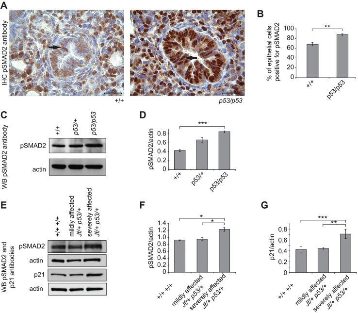
Expression of pSMAD2 and p21 in embryonic lungs. (A) Sections through E15.5 wild-type (+/+) and homozygous p53 (p53/p53) embryonic lungs stained with an anti-pSMAD2 antibody (brown stain). The nuclei were counterstained with haematoxylin. In both wild-type and mutant epithelial cells of the airways, pSMAD2 is predominantly nuclear (arrow). Scale bars: 20 μm. (B) Comparisons of the percentage of cells positive for pSMAD2 in wild-type (+/+) and p53 homozygote (p53/p53) airways. In total, 260 epithelial cells in wild types and 256 epithelial cells in p53 homozygotes were counted from airways from different regions for three embryos from each genotype. (C) Equal amounts of protein lysates from E15.5 wild-type (+/+), heterozygous (p53/+) and homozygous (p53/p53) lungs were subjected to PAGE, transferred and probed with an anti-pSMAD2 antibody. The antibody detected one main band at about 55 kDa. (D) Comparisons of pSMAD2 levels across genotypes after normalizing with an anti-actin antibody. The results presented in the graph are from three independent experiments. (E) Equal amounts of protein lysates from E15.5 wild-type (+/+ +/+) and mildly and severely affected double-mutant (Jf/+ p53/+) lungs were subjected to PAGE, transferred and probed with anti-pSMAD2 and -p21 antibodies. (F,G) Comparisons of pSMAD2 (F) and p21 (G) levels across genotypes. Data represents the analysis of four individual embryos from wild-type (+/+ +/+), and mildly and severely affected double heterozygote (Jf/+ p53/+) lungs (based on the histological observation, without and with cleft palate, respectively) after normalizing with an anti-actin antibody. Bars: standard error of mean (s.e.m.). *P<0.05, **P<0.01 and ***P<0.001. WB, western blot.

