Fig. 2.
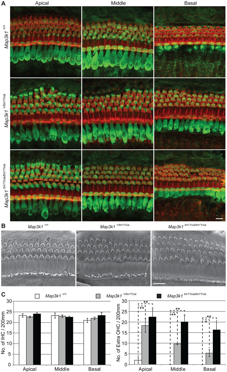
Map3k1tm1Yxia mutant mice have supernumerary outer hair cells (OHCs). (A) Whole-mount preparation of the OC from P12 mice labeled with myosin VIIa (green) and phalloidin (red). In contrast to three rows of OHCs observed in wild-type mice, Map3k1tm1Yxia mutant mice have an extra row of OHCs along the length of the cochlea, whereas sparse patches of a fourth row of OHCs was observed in Map3k1tm1Yxia heterozygous mice. Scale bar: 10 μm. (B) Scanning electron micrographs of P14 mice revealed characteristic polarized ‘V’-shaped stereocilia bundles at the apical surface of supernumerary OHCs present in Map3k1tm1Yxia heterozygous and homozygous mutant mice. Scale bar: 10 μm. (C) Quantitation of inner hair cells (IHCs) and OHCs in control and Map3k1tm1Yxia mice at P12. For quantification purposes, organ of Corti (OC) were isolated from four wild-type and Map3k1tm1Yxia mutant mice each and hair cells were counted in the apical middle and basal coil regions. No significant difference was observed in the IHC number. Statistically significant (*P<0.05, **P<0.01) increases in the OHC number were observed in the Map3k1tm1Yxia homozygous mutant and heterozygous mice, with a gradient from apex to base (mean±s.e.m.).

