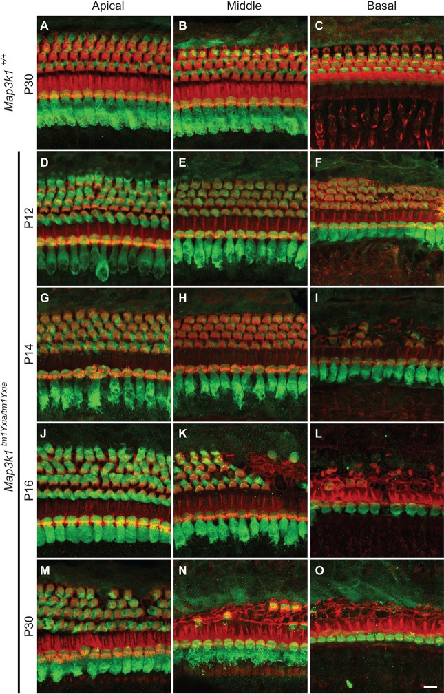
Fig. 5.
Outer hair cells (OHCs) in Map3k1tm1Yxia mutant mice degenerate as early as P14. Maximum intensity projections of confocal Z-stacks of whole-mount cochleae labeled with the anti-myosin-VIIa antibody (green) and phalloidin (red) are shown. (A-C) Representative images from the apical, middle and basal turns of the organ of Corti (OC) of a wild-type control mouse at P30. (D-O) Images of the OC from the three turns of the cochlea of Map3k1tm1Yxia mutant mice at P12 (D-F), P14 (G-I), P16 (J-L) and P30 (M-O). The hair cells appear to have normal development and morphology at P12 in the apical and middle cochlear turns in Map3k1tm1Yxia mutant mice. Initial signs of OHC degeneration are evident in the basal turn (F). At P14, obvious degeneration of OHCs was observed in Map3k1tm1Yxia mutant mice. (K,L) Severe OHC degeneration can be observed by P16 in the middle (K) and basal (L) turns. The OHC loss progresses rapidly and, by P30, severe degeneration is evident in all three cochlear turns. In contrast, inner hair cells (IHCs) remained intact along the length of the cochlea. Scale bar: 10 μm.

