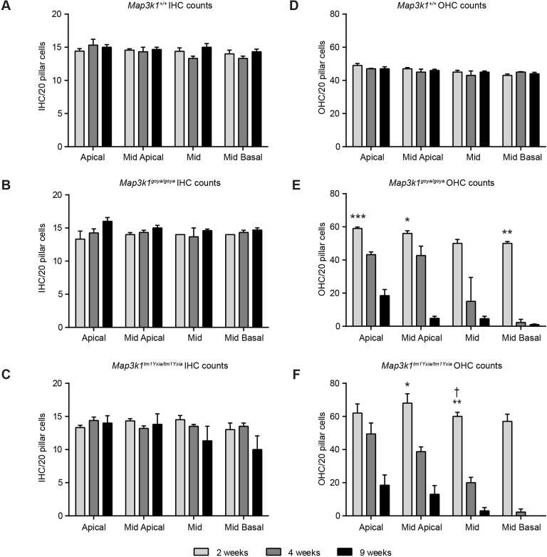
Fig. 4.
Quantification of hair-cell loss in the organ of Corti of Map3k1goya/goya and Map3k1tm1Yxia/tm1Yxia mice. (A-C) Average number of IHCs in contact with 20 pillar cells at 2, 4 and 9 weeks of age. No significant differences in IHC number were observed in wild-type (+/+; A), Map3k1goya/goya (B) and Map3k1tm1Yxia/tm1Yxia (C) mice, although, by 9 weeks of age, reduced numbers of IHCs were observed in some of the Map3k1tm1Yxia/tm1Yxia cochleae. (D-F) Average number of wild-type (+/+; D), Map3k1goya/goya (E) and Map3k1tm1Yxia/tm1Yxia (F) OHCs in contact with 20 pillar cells at 2, 4 and 9 weeks of age. t-tests were performed to compare the mean numbers of OHCs between genotypic groups at 2 weeks of age (see Materials and Methods). Map3k1goya/goya and Map3k1tm1Yxia/tm1Yxia mice have more OHCs than do wild type. This difference was significant in the apical (***P≤0.001), mid-apical (*P≤0.05) and mid-basal (**P<0.01) turns in Map3k1goya/goya. In Map3k1tm1Yxia/tm1Yxia mice, the extra number of OHCs was significantly higher than in wild type in the mid-apical (*P<0.05) and mid (**P<0.01) turns. In the mid turn of Map3k1tm1Yxia/tm1Yxia cochleae, significantly more OHCs were observed than in Map3k1goya/goya (†P<0.05); however, there were no other obvious differences between the mutant alleles. By 4 weeks of age, nearly all Map3k1goya/goya and Map3k1tm1Yxia/tm1Yxia OHCs were missing in the mid-basal turn and, in the mid turn, we observed variable levels of OHC loss in Map3k1goya/goya cochleae, and substantial loss in Map3k1tm1Yxia/tm1Yxia cochleae. In the mid-apical and apical turns, OHC loss was evident but not to the extent of the mid and mid-basal turns. By 9 weeks of age the majority of OHCs were missing in the mid-basal and mid turns of both Map3k1goya/goya and Map3k1tm1Yxia/tm1Yxia cochleae, and severe loss was seen in the mid and mid-apical turns. No significant difference in OHC numbers were seen across the time points in wild-type cochleae. The rate of decrease in hair-cell number over time was also analysed and found to be highly significant in both homozygous mutants (see Materials and Methods and Tables S1 and S2 for P-values). Data shown are mean±s.e.m., n≥3 for genotype at each cochlear turn: *P≤0.05, **P≤0.01, ***P≤0.001.

