Fig. 6.
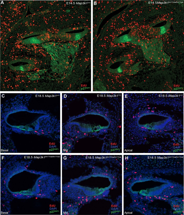
Analysis of proliferation and p27KIP1 localisation in the developing Map3k1tm1Yxia/tm1Yxia cochlea. Immunodetection of EdU-positive nuclei (red) and anti-p27KIP1 (green) and DAPI (to highlight nuclei in panels C-H). Pregnant females from timed matings were injected with EdU twice at 2-h intervals before embryos were harvested at E14.5 (A,B) or E18.5 (C-H). Localisation of p27KIP1 is unaffected in E14.5 Map3k1tm1Yxia/tm1Yxia and Map3k1+/+ cochleae (A,B). The absence of EdU-positive nuclei in the region of p27KIP1 expression indicates that the zone of non-proliferation (ZNP) is established correctly in Map3k1tm1Yxia/tm1Yxia mutant cochlea. At E18.5, we again saw no difference between the genotypes in p27KIP1 localisation, and found no evidence of proliferating cells, as denoted by EdU-positive nuclei in the cochlear duct epithelia, in any of the cochlear turns (C-H).

