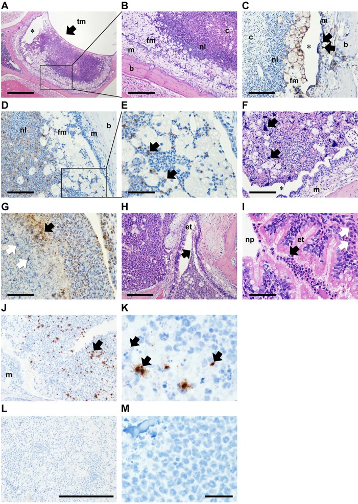
Fig. 3.
Histopathology of the Jbo/+ mouse ME 7 days after inoculation with NTHi. Composite histological features from three 12-week-old Jbo/+ mice 7 days post IN-inoculation with 106 CFU NTHi 162. (A) Haematoxylin- and Eosin-stained section of ME bulla, showing the tympanic membrane (tm; arrow). (B) Higher-magnification view (boxed area in A). Bulla fluid has a caseous core of necrotic neutrophil leukocytes (c) surrounded by viable neutrophil leukocytes (nl) and foamy macrophages (fm); inflamed ME mucosa (m) and bulla bone (b). (C) F4/80 antibody-positive foamy macrophages in bulla fluid (fm) and macrophages in the inflamed mucosa (arrows). (D,E) Cleaved caspase 3-positive apoptotic cells (arrows in E, which is a higher magnification view of the boxed area in D). (F) Two of six bulla fluids had large extracellular accumulations of Haematoxylin-positive chromatin (arrows). (G) The larger chromatin foci were histone 3 antibody negative (white arrows), but finely granular extracellular histone 3-positive material (black arrow) was scattered in the caseous core. Note normal histone staining of neutrophil and epithelial nuclei. (H,I) Neutrophils in Eustachian tube (et) lumen adjacent to the bulla opening (H) and at the nasopharynx (np) junction (I; black arrows), neutrophil leukocyte infiltration in ET submucosa (white arrows). (J,K) Jbo/+ mouse inoculated with NTHi 162sr, probed with B-HInfluenzae-NTHi375-16SrRNA oligonucleotide (Advanced Cell Diagnostic) and visualized using HRP and DAB. Bulla fluid exudates have chromogen deposits (arrows) that appeared as scattered punctate signals (∼1 μm) or irregular aggregates (10-20 μm greatest dimension); the mucosal margin is marked (m). (L,M) NTHi hybridization signals were absent in non-inoculated Jbo/+ control mice. A positive control probe (Ppib) for mouse RNA integrity showed punctate signals in mucosal epithelium and bulla fluid cells, but no signal was obtained with a negative control probe (bacterial DapB gene); data not shown. Scale bars: 500 µm in A, 200 µm in B,H, 100 µm in C,D,F and 50 µm in E,G,I. In A,C,F, asterisk indicates an artefactual cleft caused by histological processing. (J,L) Magnification ×200; scale bar: 200 µm. (K,M) ×1000 oil immersion; scale bar: 20 µm.

