Fig. 4.

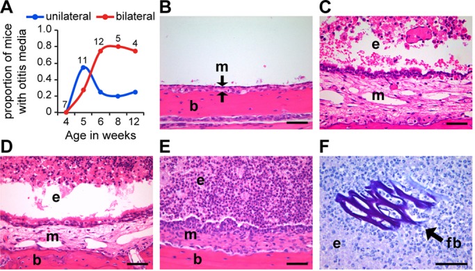
Jbo/+ mice develop OM in GF conditions. (A) Time course of the proportion of Jbo/+ GF mice with unilateral or bilateral OM (the number of mice is indicated alongside each time point). (B-F) Histological analysis of Jbo/+ mouse ME. (B) Haematoxylin- and Eosin-stained sections of non-inflamed ME of a 4-week-old GF Jbo/+ mouse with an air-filled bulla space, thin mucosa (m) indicated between arrows, supported by underlying bulla bone (b). (C) Inflamed ME of a 4-week-old SPF Jbo/+ mouse with exudate (e) in the bulla space and thickened mucosa (m). (D,E) Inflamed ME of GF Jbo/+ with bulla exudate (e) and thickened mucosa (m) at 5 (D) and 8 weeks of age (E). (F) ME exudate in a 12-week-old GF Jbo/+ mouse contains plant-based foreign body (fb) in bulla exudate (periodic acid-Schiff-stained section). Scale bars: 50 µm in B-F.
