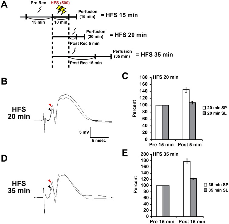
Fig. 1.
High frequency stimulation (HFS)-induced L-LTP potentiates spike amplitude and field EPSP slope in the dentate gyrus. (A) Experimental schedule for this study. Sampling was performed after transcardial perfusion of paraformaldehyde. The perfusion began 15, 20 or 35 min after the start of HFS (10 min). (B-E) Data obtained at 20 min (B,C) and 35 min (D,E) under HFS conditions. (B,D) Typical waveforms pre- and post-HFS are indicated by black and red arrowheads, respectively. (C,E) Relative changes in population spike amplitude (SP) and field EPSP slope (SL) after HFS. Data from each condition were obtained from three animals. Data shown as mean±s.e.m.

