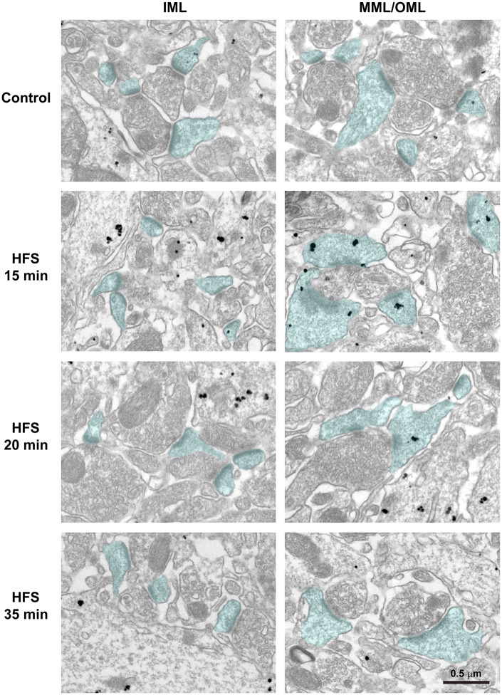
Fig. 4.
Immunoelectron microscopic observation of ribosomal protein S6 (rpS6) distribution after high frequency stimulation (HFS). Immunohistochemistry was performed with hippocampal dentate gyrus sections from the contralateral hemisphere and the ipsilateral hemisphere of HFS 15 min, 20 min, or 35 min condition. Micrographs of inner molecular layer (IML) (left panels) and middle/outer molecular layers (MML/OML) (right panels) are shown. Nanogold particles indicate rpS6 signals. Spines containing postsynaptic densities are blue. Scale bar: 0.5 µm.

