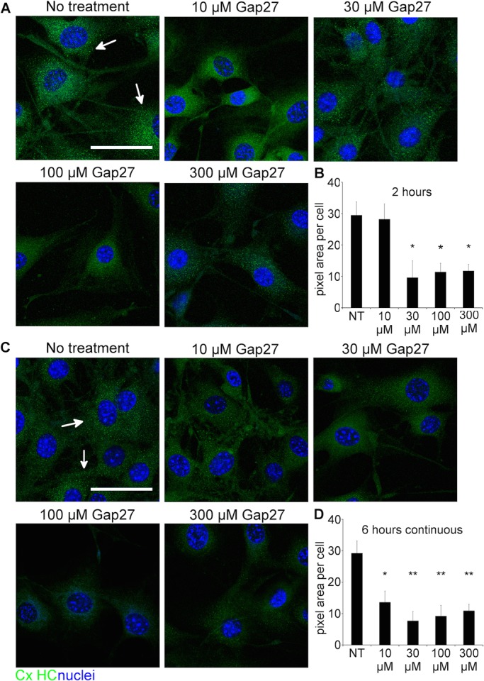
Fig. 2.

Gap27 reduced CxHC protein levels in normal 3T3 fibroblasts. (A,C) Representative images of 3T3 cells with no treatment (NT), or treated with increasing doses of Gap27 for (A) 2 h or (C) 6 h. Cells were immunolabelled for CxHC (green, white arrows) and nuclei (blue) identified by Höechst. (B,D) CxHC labelling intensity was quantified at (B) 2 h and (D) 6 h. Data are means±s.e.m. (n=3). *P<0.05, **P<0.01 compared with NT, one-way ANOVA. Scale bar=50 µm.
