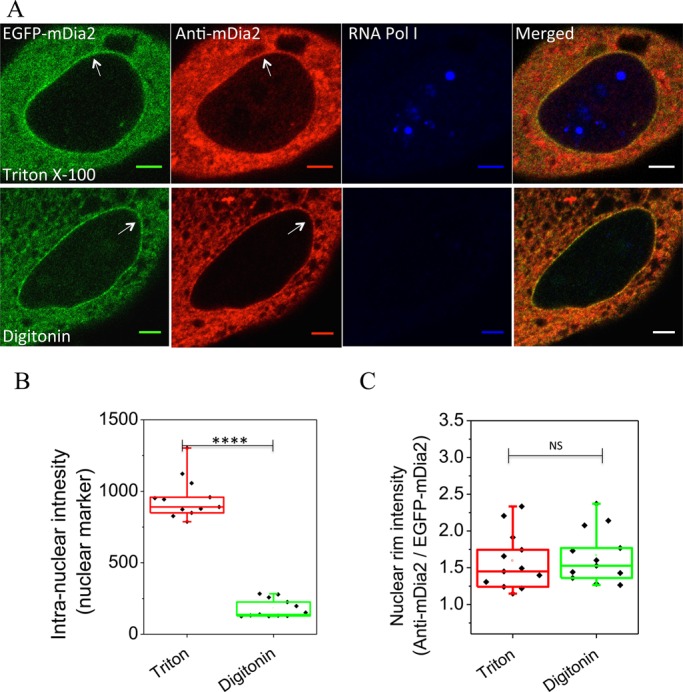
Fig. 2.

mDia2 localization at the cytoplasmic side of the nuclear envelope. (A) Immunostaining of mDia2 (red) and an intra-nuclear protein PRAF1, a component of RNA polymerase I (RNA Pol I, blue), in EGFP-mDia2 (green) expressing cells permeabilized by either Triton X-100 (upper) or digitonin (lower). Superimposed images are shown on the right. Note that digitonin does not permeabilize nuclear membranes. Arrows indicate enrichment of mDia2 at the nuclear rim. Scale bars: 5 μm. (B) Quantification on intensity of PRAF1 inside the nucleus in cells permeabilized by either Triton X-100 or digitonin. (C) Quantification on intensity of mDia2 immunostaining (normalized by that of EGFP-mDia2) at the nuclear rim in the same cells as quantified in (B). ****P<0.0001; NS, P>0.05; two-tailed unpaired Student's t-test.
