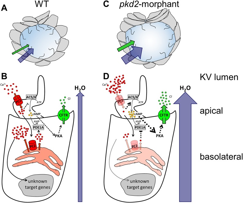
Fig. 5.
The zebrafish KV as a model organ for CFTR stimulation in ADPKD cysts. (A,B) In WT embryos, the KV inflation is ensured by the CFTR-mediated transport of Cl− and by the subsequent movement of water towards the organ lumen (A). In KV epithelial cells (B), Polycystin-2 (PC2) located at the cilia membrane allows the entrance of Ca2+ when stimulated by the luminal fluid-flow. This ciliary wave activates the Ca2+ release from the ER pools, in a Polycystin-2-dependent manner, initiating a Ca2+-signaling of unknown effectors. Through inhibition of adenylyl cyclases 5 and 6 (AC5/AC6) and activation of phosphodiesterase 1A (PDE1A), the Ca2+ transients maintain the basal intracellular levels of cAMP required for the normal rate of CFTR activity. (C,D) Mimicking ADPKD cysts, the pkd2-knockdown enhances CFTR-mediated ion and fluid secretion into the KV, resulting in its significant enlargement (C). The reduced Ca2+ oscillations are expected to activate AC5/AC6 and inhibit PDE1A, raising the intracellular levels of cAMP and, thus, driving the overstimulation of CFTR (D). Ca2+ and Polycystin-2 (red); Cl− and CFTR (green); cAMP (yellow); H2O (blue); AC5, AC6, and PDE1A (grey). Full black arrows – known activations; dashed lines and arrows – expected inhibitions/activations; line and arrow widths are proportional to the expected level of activation.

