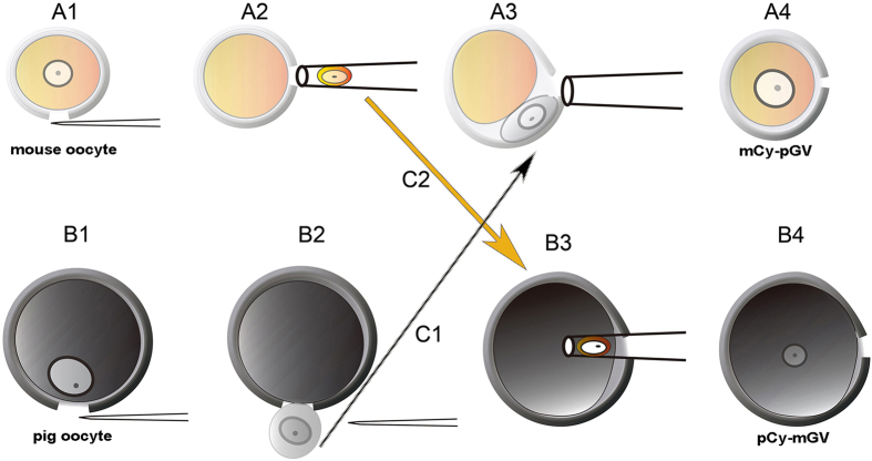
Figure 1. Procedure of GV transfer between mouse and pig oocytes.
(A1) Zona pellucida of mouse oocytes was cut with a sharp glass needle. (A2) Mouse GV was aspirated by a blunt-tip micropipette with an inner diameter of about 20 μm. (A3) Pig GV was transferred to the perivitelline space of enucleated mouse oocyte by a blunt-tip micropipette with an inner diameter of about 25 μm. (A4) Pig GV was fused to the mouse cytoplast by electric fusion. (B1) Zona pellucida of mouse oocyte was cut by using pressure with a sharp glass needle. (B2) Pig GV was squeezed out by pressing the zona pellucida. (B3) Mouse GV was directly injected into pig cytoplast by a blunt-tip micropipette with an inner diameter of about 20 μm with a piezo unit. (B4) A pCy-mGV oocyte was reconstructed with pig cytoplast and mouse GV. (C1) Pig GV was transferred to the micro-manipulation droplet with enucleated mouse oocyte. (C2) Mouse GV was transferred to the micro-manipulation droplet with enucleated pig oocyte.

