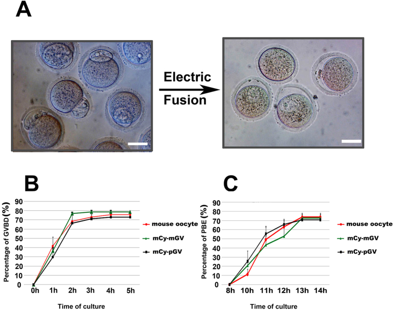
Figure 3. In vitro maturational time course is similar between mCy-pGV oocytes and mouse oocytes.
(A) Pig GV was fused to the mouse enucleated oocyte by electric fusion. (B) Percentages of GVBD in mouse oocytes (0 h: 0%; 1 h: 41.61 ± 11.35% 2 h: 68.51 ± 9.84%; 3 h: 73.11 ± 6.68%; 4 h: 75.53 ± 2.57%;5 h: 75.53 ± 2.57%), mCy-mGV oocytes (0 h: 0%; 1 h: 36.62 ± 0.24%; 2 h: 76.99 ± 3.17%; 3 h: 78.74 ± 1.38%; 4 h: 78.74 ± 1.38%; 5 h: 78.74 ± 1.38%) and mCy-pGV oocytes (0 h: 0%; 1 h: 29.81 ± 6.05%; 2 h: 66.37 ± 12.46%; 3 h: 71.64 ± 8.91%; 4 h: 72.92 ± 6.82%; 5 h72.91 ± 6.82%) were observed each hour from 0 to 5 h of in vitro culture. (C) Percentages of mouse oocytes (8 h: 0%; 10 h: 10.57 ± 4.41%; 11 h: 49.17 ± 13.72%; 12 h: 63.10 ± 8.22%; 13 h: 73.69 ± 3.04%; 14 h: 73.69 ± 3.04%), mCy-mGV oocyte(8 h: 0%; 10 h: 20.88 ± 3.76%; 11 h: 43.46 ± 8.90%; 12 h: 52.65 ± 5.84%; 13 h: 72.71 ± 5.28%; 14 h: 72.71 ± 5.29%) and mCy-pGV oocytes (8 h: 0%; 10 h: 25.56 ± 11.16%; 11 h: 55.27 ± 8.33%; 12 h: 65.65 ± 3.71%; 13 h: 70.78 ± 5.42%; 14 h: 70.78 ± 5.42%) with first polar body extrusion were detected 8 h, 10 h, 12 h, 13 h, and 14 h of in vitro culture (bar = 30 μm).

