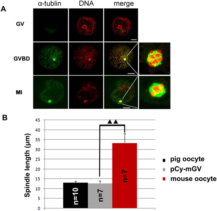
Figure 6. Spindle size of pCy-mGV oocytes.

(A) pCy-mGV oocytes at GV, GVBD and MI stages were stained with PI and anti-α-tubulin antibody. Normal chromosome condensation could be detected in GVBD stage oocytes. A small spindle was assembled in the pCy-mGV oocytes. (B) Spindle size of the MI stage pCy-mGV oocytes were12.80 ± 1.27 μm, significantly small than that of mouse oocytes (33.33 ± 4.80 μm) (p < 0.05), but similar to that of pig oocytes (13.17 ± 0.74 μm) (bar = 30 μm).
