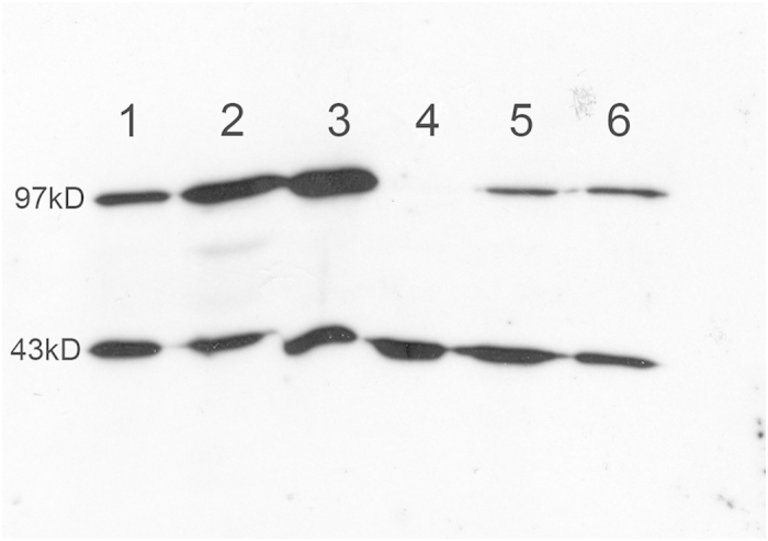
Figure 6. Western blot analysis using an anti-active β-catenin antibody.

Western blot analysis on three APAs with CTNNB1 mutation (1–3), one APA without known mutation (4) and two APAs with KCNJ5 mutation (5–6). Top row shows signal for active β-catenin (97 kD), lower row shows actin signal (43 kD).
