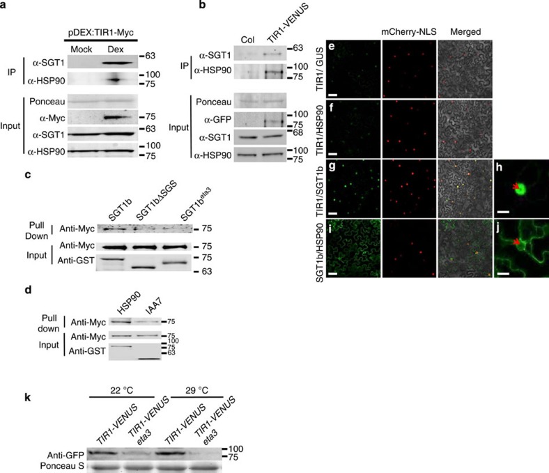
Figure 5. TIR1 is in a complex with HSP90 and SGT1.
(a,b) Immunoprecipitation (IP) assays were performed using 7-day-old pDEX:TIR1-Myc (a) or pTIR1:gTIR1-VENUS (b) seedlings. TIR1, SGT1b and HSP90 were detected with the appropriate antibodies. (c) In vitro pull-down assay with GST-tagged SGT1b, SGT1bΔSGS and SGT1b(eta3). TIR1-Myc was synthesized in a TNT extract. (d) In vitro pull down with GST-tagged HSP90.2 and TIR1-Myc. GST-IAA7 was used as positive control. (e–j) YFP-fusion genes were introduced into N. benthamiana leaves by infiltration with A. tumefaciens. (e) nYFP-TIR1 and cYFP-GST (negative control). (f) nYFP-TIR1 and cYFP-HSP90. (g) nYFP-TIR1 and cYFP-SGT1b. (i) nYFP-SGT1b and cYFP-HSP90. (h,j) Enlarged images of single nuclei in (g) and (i) are shown, respectively. The arrows indicate the nucleolus. mCherry-NLS highlights nuclei. (k) Seven-day-old seedlings were transferred to 29 °C or kept at 22 °C for an additional 24 h before the tissue was harvested for protein blots. Scale bars, 50 μm (e,f,g,i) and 10 μm (h,j).

