Figure 5.

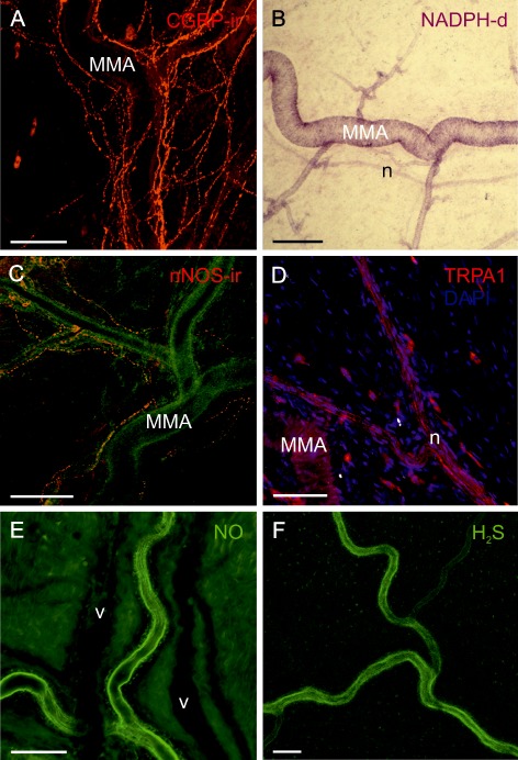
Histochemical demonstration of components of HNO‐CGRP signalling in the dura mater; size bars 100 μm. (A) Confocal immunofluorescence image of CGRP‐immunoreactive nerve fibres densely surrounding the MMA and its branches. (B) Transmission light microscopic image showing NADPH‐diaphorase staining of the MMA and some arterioles up to the transition into capillaries, indicative for the production of NO. An adjacent nerve bundle (n) is weakly stained. (C) Confocal image of nerve fibres immunoreactive for nNOS surrounding the MMA, which is visible in the green background channel. (D) Confocal image showing TRPA1‐immunoreactive nerve fibres in a nerve bundle (n) adjacent to the MMA. (E) Confocal image showing arterial endothelium of the MMA stained by DAF indicating NO. Two unstained accompanying veins are visible in the background as dark shadows. (F) Confocal image showing endothelial staining of the MMA by WSP1 demonstrating the presence of H2S.
