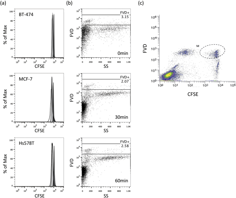
Figure 2. Staining strategy of the new flowcytometric assay.
(a) Breast cancer cell lines, BT-474, MCF-7 and Hs578T, were stained with CFSE, and the intensity was then measured by flowcytometry, soon after staining (black filled histogram), after an overnight incubation (gray-filled histogram) and after 2 days of incubation (open histogram). (b) Human PBMCs were stained with FVD (1:1000 dilution, at RT for 20 min.), PI (2 μg/mL, at RT for 10 min.), or 7-AAD (5 μg/mL, at RT for 10 min.), and the dead cells (%) were then measured by flowcytometry. (c) Data obtained with the new flowcytometric analysis method. CFSE + cells are target cells, and FVD + cells are dead cells, such that dead target cells are represented by the CFSE + FVD + area.

