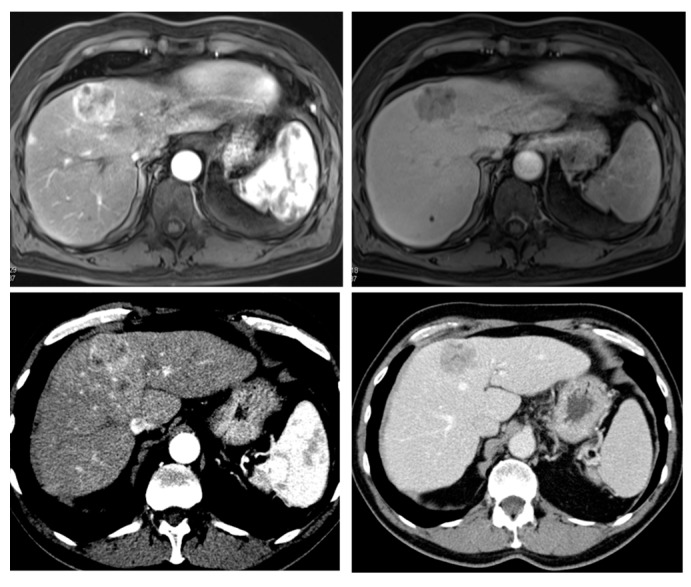
Figure 1.
Radiological hallmarks of hepatocellular carcinoma (HCC). Typical vascular pattern of HCC as observed in a 67-year old patient with histology proven HCC. Liver lesion in the right hepatic lobe observed in a cirrhotic patient. The lesion is presenting a typical HCC vascular pattern with arterial hyperenhancement (left images) and venous wash-out (right images) visible both in magnetic resonance imaging (MRI) (upper row) and computed tomography (CT) (lower row).

