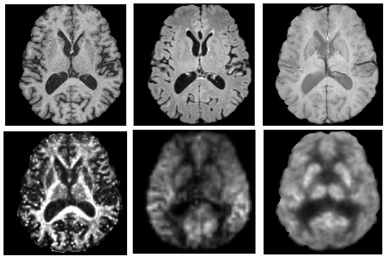
Figure 2.
PET and MR images of a 77-year-old male injected with 198 MBq of 18F-flurodeoxyglucose (FDG) while patient was lying supine in a Siemens Biograph mMR PET-MR hybrid scanner (Siemens Healthcare, Erlangen, Germany). A 60 min dynamic PET scan was acquired during multispectral MR imaging. These images illustrate the capacity to perform multi-parametric mapping in a single session simultaneously, to improve characterization of neuropsychiatric conditions. Axial images are from (top) left to right: T1-weighted (MRPAGE) for tissue-specific volumetric measurements, T2-weighted (FLAIR) for assessment of white matter lesions, and susceptibility-weighted imaging for detection of microbleeds and cerebral amyloid angiopathy; (bottom) left to right: fractional anisotropy image from diffusion tensor imaging for quantification of white matter structural integrity, perfusion weighted-imaging (ASL) for hemodynamic measurements and a PET-18F-FDG glucose consumption image. Images are presented with permission from patient and are courtesy of Lawson imaging, Lawson Health Research Institute (LHRI), London, ON, Canada.

