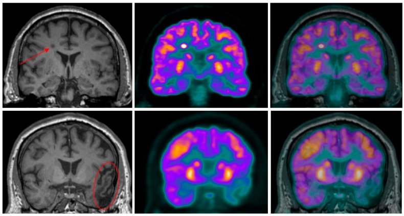
Figure 3.
Coronal high-resolution T1-weighted MR (left) and PET (center) images of two pulmonary carcinoma patients scanned at LHRI, London, Ontario, Canada using a Siemens Biograph mMR PET-MR hybrid scanner. The PET-18F-FDG and MR images were acquired simultaneously and, the PET was later superimposed onto the MR images (right). To illustrate the synergistic effect of PET’s high sensitivity and MR’s high spatial resolution to improve the specificity of characterizing neuropsychiatric disorders, two case studies are presented. (Top) row: A focal area of increased FDG-PET uptake in a metastatic lesion can be seen on the top row images that is well defined when the PET image is overlayed onto the MR image; (Bottom) row: Areas of hypometabolism in PET-18F-FDG images, a result of prior traumatic brain injury, are delineated in the fused PET and MR images. Images are presented with permission from patients.

