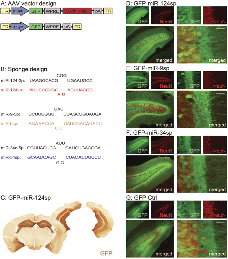
Figure 2. miRNA inhibition using AAV-miRNA sponges.
(A) AAV-miRNA sponges were expressed in order to inhibit individual miRNA families. These vectors express a GFP reporter under the influence of a synapsin promoter (p-syn) with (upper panel) or without (lower panel; GFP Ctrl) the presence of eight imperfectly complementary target sites for a specific miRNA, all within inverted terminal repeats (ITRs) of an AAV2/5 vector. (B) The design of the miR-124sp, miR-9sp and miR-34sp sequences in comparison to the endogenous miRNA. (C–G). (C) DAB immunohistochemistry, (D–G) fluorescence immunohistochemistry. Injection of AAV2/5 pseudotyped vectors into the hippocampus (three locations per hemisphere), resulted in the expression of the GFP reporter in a majority of hippocampal neurons of the dentate gyrus in the presence of a miR-124sp sequence (C–D), a miR-9sp sequence (E), a miR-34sp sequence (F), or without such a sequence (G). Scale bars 100 μm (D–G); (left merged panels) and 20 μm (D–G); (right merged panels).

