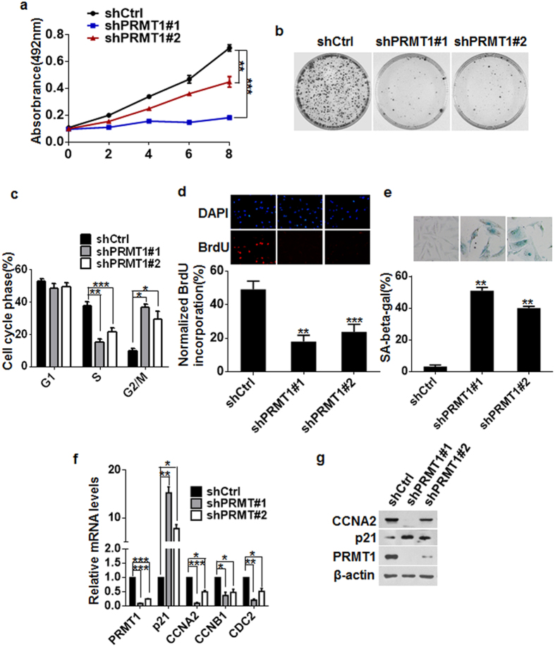
Figure 4. Knockdown of PRMT1 arrested breast cancer cell growth in G1 tetraploidy and induced cellular senescence.
(a) Effect of PRMT1 knockdown on cell proliferation in MDA-MB-231 cells was assessed by MTT assays at different time points as indicated. Data are the mean ± SD of five replicates per experiment. (b) Cell proliferation was examined by colony formation assays in MDA-MB-231-shPRMT1#1/#2 and shCtrl cells. (c) Cell cycle analysis of MDA-MB-231-shPRMT1#1/#2 and shCtrl cells at day 4 after lentivirus infection. Percentages of subpopulation of cells at different cell cycle phases based on triplicate experiments. (d) Representative immunofluorescence images of BrdU incorporation in MDA-MB-231-shPRMT1#1/#2 and shCtrl cells. Lower panel represents the mean number of cells per field ± SD based on cell counts from five randomly chosen fields. (e) MDA-MB-231-shPRMT1#1/#2 and shCtrl cells were subjected to SA-β-Gal staining to determine the percentage of the senescent population (lower). Top panel represents images of SA-β-Gal staining. (f,g) Expression of p21 and G2/M associated proteins was analyzed by qRT-PCR (f) and western blotting (g). All experiments were repeated at least three times. Error bars, mean ± SD, *P < 0.05, **P < 0.01, ***P < 0.001.

