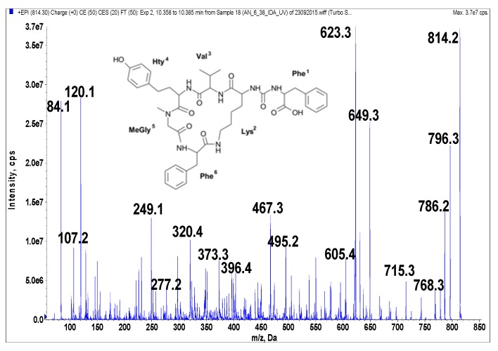
Figure 5.
Schematic structure and mass fragmentation spectrum of anabaenopeptin AP 813. The structure of the peptide (Phe + CO[Lys + Val + Hty + MeGly + Phe]) was deduced on the basis of the following fragments: 796 [M + H − H2O], 786 [M + H − CO], 768 [M + H − CO − H2O], 715 [M + H − Val], 649 [M + H − Phe − H2O], 623 [M + H − (CO + Phe)], 605 [M + H − (CO + Phe) − H2O], 495 [Phe + MeGly + Hty + Val + H], 467 [Phe + Lys + CO + Phe + H], 396 [Hty + MeGly + Phe + H], 373 [M + H − Phe − (Hty + Val) − H2O], 320 [M + H − Phe − (Val + Hty + MeGly + Phe)], 277 [Hty + Val + H], 249 [Hty + MeGly + H], 120 Phe-immonium ion, 107 [CH2PhOH], 84 Lys-immonium ion.

