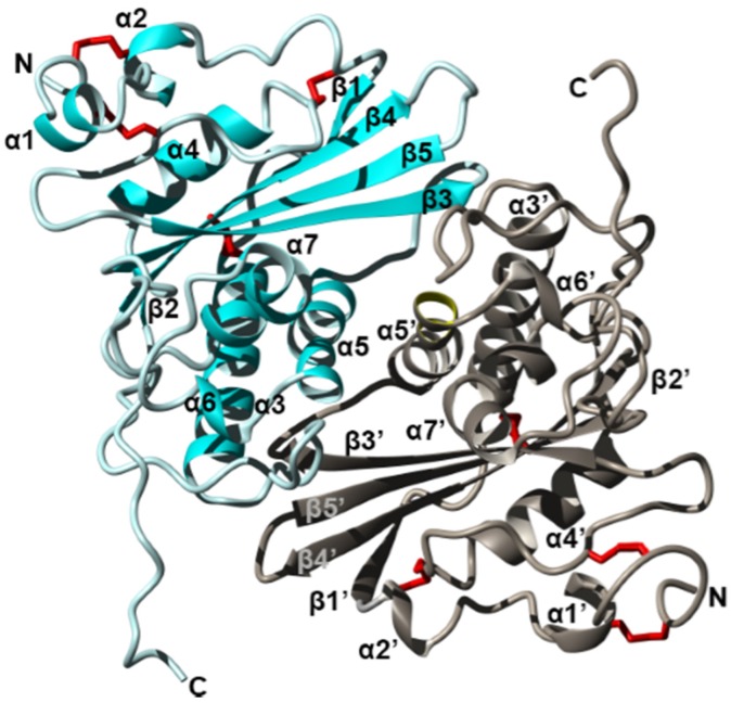
Figure 3.
Crystal structure of the fire ant (Solenopsis invicta) venom allergen Sol i 3 dimer (PDB accession 2VZN). Ribbon diagram revealing the overall structure of each monomer which contains seven helices (α1–α7) and five beta strands (β1–β5), arranged as three stacked layers, giving rise to an α–β–α sandwich. Two units (cyan and silver) form a dimer by non-disulfide bonds involving symmetrical residues in helix α5 and α5′. Disulfide bridges are shown in red, and N and C termini are labelled. Figure modified from Padavattan et al. Redrawn from [125].

