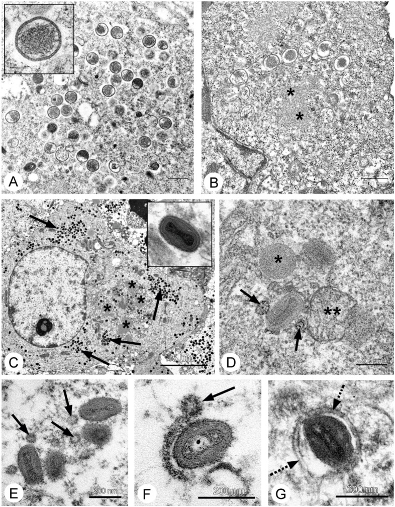
Figure 3.
Replication of the L-IVP strain of VACV in cells of human A431 carcinoma (A,C,E–G) and murine Ehrlich ascitic carcinoma (B,D) cells. Photos A,B show the formation of immature virus particles, the insert in A shows a virion at high magnification, the asterisks show a viroplasm. Photo C shows infected cell containing viroplasm (*) and accumulations of mature virions (shown by arrows), the insert for C shows a mature virion at large magnification. Photos D–G show sequential steps of formation of “enveloped” virions. The arrows show Golgi-derived vesicles, which fuse and thereby form double-membrane envelope (shown by dotted arrow). The asterisk on photo D shows immature virion, the double asterisks show mitochondria.

