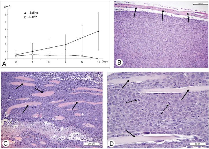
Figure 4.
Changes of murine Ehrlich solid carcinoma volume after injection of the L-IVP strain and saline (A); period after virus injection is shown on X-axis, tumor volume—on Y-axis. Paraffin section of A431 carcinoma showing capsule (shown by arrows) and smooth outline (B); and Ehrlich solid carcinoma (C,D). Arrows show fragments of muscle tissue, asterisks show necrotic zone, dotted arrows show mitoses. Hematoxyline and eosin staining.

