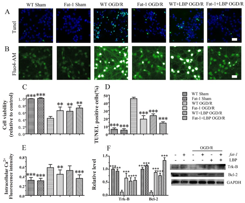
Figure 4.
LBP and endogenous ω-3 PUFAs significantly prevent OGD/R-induced neuronal apoptosis respectively via intracellular Ca2+ handling or neurotrophic pathway activation: (A) TUNEL staining; (B) fluorescent micrographs showing intracellular Ca2+ levels as stained by the Fluo4-AM dye; (C) statistic of cell viability; (D) statistic of TUNEL positive cells; (E) results of relative fluorescence intensity analysis of intracellular Ca2+; and (F) expression levels of Trk-B and Bcl-2 measured by Western blot. Data are presented as mean ± SEM, ** p < 0.01, *** p < 0.001 indicate significant difference compared with the WT OGD group; p < 0.05, p < 0.01 indicates significant difference compared with the WT DHA + LBP group (t-test). Scale bar: 50 µm.

