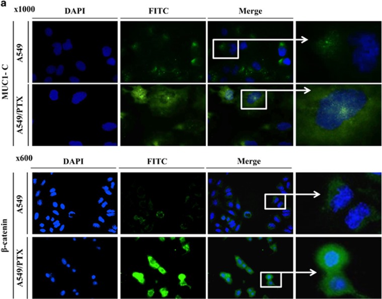
Figure 2.
Immunofluorescence of MUC1-C and β-catenin in A549/PTX. (a) A549 and A549/PTX cells were fixed and assayed with immunofluorescence. DAPI nuclear staining is shown at low magnification; the boxed regions are at a higher magnification. Fluorescence images of MUC1-C (magnification, × 1000) and β-catenin (magnification, × 600).

