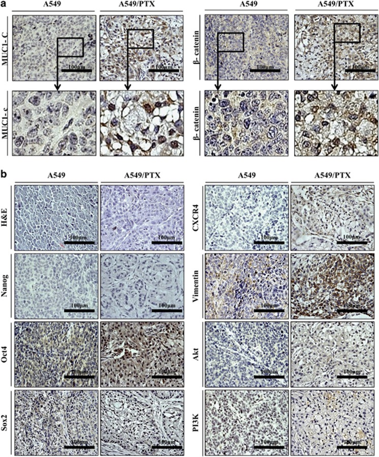
Figure 4.
Immunohistochemical staining of A549/PTX cell tumors. (a) Immunochemistry images of A549 and A549/PTX tumors using respective antibodies specific to MUC1-C and β-catenin. The boxed regions are at a higher magnification. (b) Immunohistochemical analysis of stemness genes, EMT markers and survival factors (magnification, × 400).

