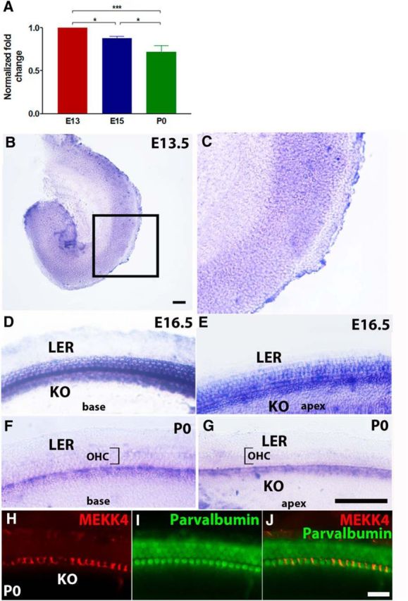
Figure 1.

Spatiotemporal expression pattern of MEKK4 in the mouse inner ear. A, qPCR-based analysis reveals developmental changes in the levels of MEKK4 expression within WT mouse inner ears. Although MEKK4 is abundantly expressed in the developing inner ear as early as E13, its expression steadily decreased from E13 to P0. B–G, Whole-mount in situ hybridization for MEKK4 at E13.5 (B, C), E16.5 (D, E), and P0 (F, G) shows that MEKK4 is initially expressed broadly within organ of Corti; and by E16.5, expression is seen in developing HCs and supporting cells. By P0, expression becomes restricted to inner phalangeal cells within the medial domain of the organ of Corti. H–J, Immunolabeling of P0 cochlea using antibodies against MEKK4 (red) and parvalbumin (green) demonstrates the expression of MEKK4 in the soma of inner phalangeal cells. The results represent an average of six samples at each developmental age group. Data are mean ± SEM. KO, Kolliker's organ; LER, lesser epithelial ridge. Scale bars: (in B) B, C, 50 μm; (in G) D–G, 20 μm; (in J) H–J, 20 μm.
